

Clinical Lead
Meva Clinic Plastic Surgery Care Team
Otoplasty (Prominent Ear Surgery)
Reshapes prominent ears to support more natural and balanced facial proportions.
Meva Clinic Plastic Surgery Care Team
Patient Care Information
Patient Care Information
"Our clinical approach prioritizes patient safety, individualized treatment planning, realistic expectations, and long-term follow-up throughout the international care journey."
About This Procedure
Why Choose Meva Clinic?
Reshapes prominent ears to support more natural and balanced facial proportions.
Ears that stand out from the head, or differ from one another, are a common reason for self-consciousness in adults. Otoplasty in Istanbul reshapes the ear cartilage through a discreet incision behind the ear, so the scar sits in the natural fold. Structural sutures hold the new position. A free online pre-assessment from photos is a sensible first step. At the consultation, the surgeon looks at which part of the ear causes the prominence, whether the fold is missing or the bowl of the ear is deep, and plans accordingly. The procedure is done under local anaesthesia or sedation, without a hospital night. You stay about 4 hotel nights and can usually return to office work after 3 days. For a profile-focused change, see rhinoplasty.
Patient Care Information
Meva Clinic Plastic Surgery Care Team
Aesthetic & Body Contouring Pathway Guidance
This content is maintained by Meva Clinic’s Clinical Care Team for patient guidance and clinical transparency. Final treatment suitability, operating physician assignment, surgical planning, and medical decisions are confirmed only after medical history review, in-person consultation, diagnostic tests, anesthesia evaluation, and doctor availability.
Is This Treatment for Me?
Prominent ears
Asymmetrical ears
Children from about age 5–6 and adolescents, with parental consent
Adults self-conscious about their ears
Comprehensive International Patient Care Plan
Private airport transfer according to the confirmed care plan
Bosphorus-view partner hotel, according to the confirmed care plan
Dedicated patient support during the confirmed care pathway
Aftercare guidance or materials according to the confirmed treatment plan
Follow-up guidance according to individual medical recommendations
The Clinical Procedure
Cartilage reshaping through a discreet posterior incision.
Ear Assessment and Photos
The surgeon examines both ears to see what causes the prominence, a missing fold, a deep ear bowl or both, and takes standardised photos for planning.
Local Anaesthesia, Optional Sedation
The ears are numbed with local anaesthesia, and light sedation can be added for comfort. No hospital night is needed.
Incision Behind the Ear
A small incision is made on the back of the ear, so the scar later lies in the natural crease between ear and head.
Cartilage Reshaping
The cartilage is shaped and held with fine non-absorbable internal sutures to create the missing fold; if the ear bowl is deep, it is set back closer to the head.
Symmetry Check and Closure
Both ears are compared for symmetry, then the skin is closed with fine sutures hidden in the ear crease.
Padded Dressing and Headband
A soft padded dressing protects the ears for the first days, followed by an elastic headband that supports the new position.
Recovery & Transformation Journey
Phase 1: Days 0–2
You rest at the partner hotel on the same day. The ears feel tight and tender; oral pain relief usually keeps this comfortable. Keep the dressing dry and sleep with your head raised.
Phase 2: Days 3–7
The dressing is removed at the check-up and replaced by a headband. Office work is usually possible from about day 3, and you fly home after the final check, around 4 hotel nights.
Phase 3: Weeks 2–6
Swelling and bruising fade. The headband is typically worn day and night at first, then only at night for several weeks; avoid contact sports and pressure on the ears. Your surgeon will confirm the exact schedule.
Phase 4: Months 2–6
Residual swelling settles and the scar behind the ear softens and fades. The final shape is usually assessed at around 3–6 months, with remote follow-up by photos.
The Meva Advantage
Structural suture techniques designed to support stable, long-lasting ear positioning.
Medical Guidelines & Safety Protocols
Hospital-based procedures are coordinated through accredited partner hospitals, including JCI-accredited institutions where clinically appropriate. Below are the clinical guidelines, safety protocols, and medical reference documentation for this procedure.
Indicated / Suitable For
- Children, adolescents and adults whose ears stand out from the head because of a missing antihelical fold, a deep conchal bowl or both
- Patients with noticeable asymmetry in the position or projection of the two ears
- Children from about 5–6 years of age, when the ear has reached most of its adult size, and adolescents, assessed individually and treated with the consent and presence of their legal parent or guardian
- Patients in good general health who are fit for the planned anaesthesia: usually local anaesthesia with sedation in adults, often general anaesthesia in young children
- People with realistic expectations of better-balanced ears rather than two perfectly identical ears
Contraindications / Not Suitable
- Active skin infection, eczema or inflammation around the ears or on the scalp
- A tendency to keloid or hypertrophic scarring, which requires specific discussion before surgery
- Inability to wear the protective headband as instructed or to avoid contact sports during healing
- Bleeding disorders or blood-thinning medication that cannot be safely paused
- Ear shape problems caused by other conditions (such as a major congenital ear deformity) that need a different, reconstructive approach
Pre-Operative Evaluation & Diagnostic Checks
Ear Analysis: Measurement of ear projection and identification of the cause on each side (missing fold, deep conchal bowl, prominent earlobe).
Cartilage and Skin Assessment: Checking cartilage stiffness, skin quality behind the ear and any history of abnormal scarring.
Standardised Photography: Front, back and side photos to plan symmetry and to compare results at follow-up.
Health, Anaesthesia and Consent Review: Basic blood tests when indicated and a review of medicines that affect bleeding. Adults usually have local anaesthesia with sedation; for children, the surgeon and anaesthetist decide the anaesthesia (often general anaesthesia in young children) and the legal parent or guardian signs the consent.
Potential Risks & Complications
In compliance with transparent clinical reporting, Meva Clinic ensures patients are fully informed of the physiological risks associated with surgical procedures through accredited partner-hospital safety protocols where clinically appropriate.
Some ears drift partly back towards their original position, most often in the first months; secure cartilage sutures and wearing the headband as advised reduce this, and a secondary correction can be considered if needed.
Small differences between the two ears are common during healing; both sides are compared during surgery and the final position is judged once swelling has settled.
Blood collecting under the skin, causing increasing one-sided pain, usually within the first 48 hours; the dressing check allows early detection and prompt drainage.
Infection is uncommon and is treated with antibiotics; a buried suture may occasionally become visible or irritating (suture extrusion) and can be removed once the shape has stabilised.
The scar lies in the crease behind the ear and rarely thickens (hypertrophic scar or keloid). Temporary numbness and cold sensitivity of the ear usually improve over a few months.
Sharp edges or an over-corrected, pinned-back look are avoided by gradual, natural shaping of the cartilage.
Realistic Expectations & Outcomes
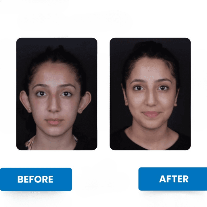
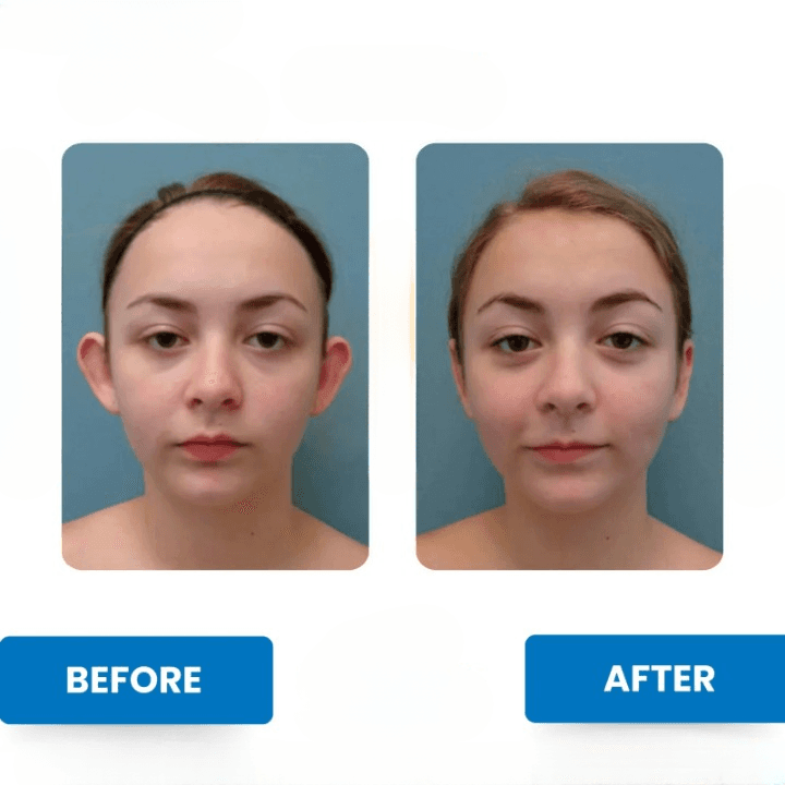
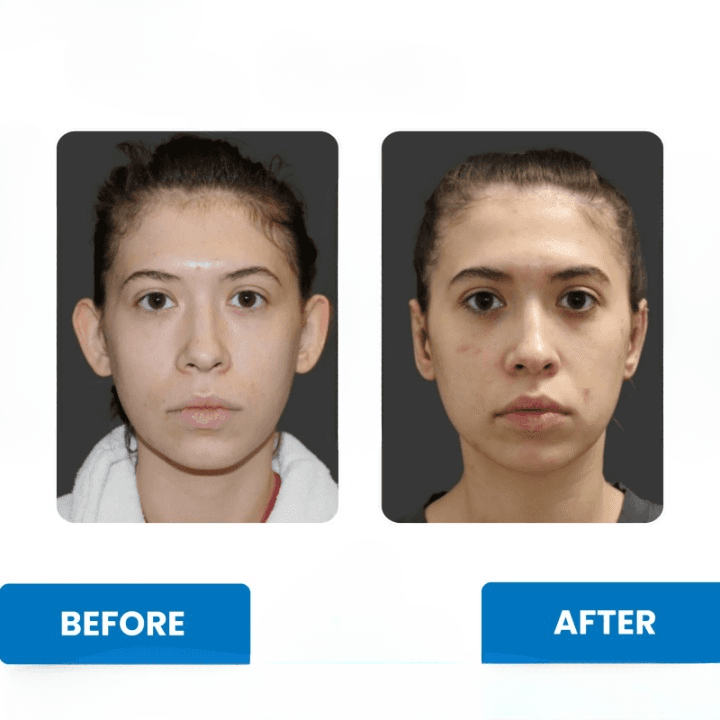
Otoplasty brings prominent ears closer to the head and recreates a natural ear fold, so the ears sit in better balance with the face. It improves position and shape; it does not substantially change ear size or make both ears perfectly identical, as ears naturally differ slightly. The ears are swollen, tender and sometimes bruised in the first week, and the headband supports the new shape while the cartilage settles. Most swelling fades over a few weeks, but mild thickness and numbness can last longer. The final shape is usually judged at around 3 to 6 months.
Revision & Follow-Up Policy
Remote follow-up with photos is planned at around 1, 3, 6 and 12 months after you return home. Because cartilage and soft tissue need several months to stabilise, partial recurrence or asymmetry is evaluated only after this period, usually from 6 months. If a secondary correction is considered, it is discussed individually with the surgeon based on objective findings. For 12 months after your treatment you can also send photos to the medical team whenever you wish and receive their feedback, and your questions are answered remotely.
Authoritative Medical References & Studies
- [1]Sadhra SS, Motahariasl S, Hardwicke JT. Complications after prominent ear correction: A systematic review of the literature. J Plast Reconstr Aesthet Surg. 2017;70(8):1083-1090.View Source
- [2]Thomson H, Gosden J, Mandal A. Otoplasty for prominent ear: A systematic review of surgical techniques. JPRAS Open. 2026;49:221-233.View Source
Medical Disclaimer: The content on this page is provided for information and education. Ear anatomy, cartilage stiffness and healing vary widely between individuals. Suitability for otoplasty, the technique used and the likely result are decided only after a personal examination by a qualified specialist plastic surgeon.
Frequently Asked Questions
Related Treatments
Piezo Ultrasonic Rhinoplasty
Advanced ultrasonic nasal reshaping using high-frequency micro-vibrations. Designed for selective bone remodeling with soft-tissue preservation to support comfortable healing; individual recovery varies.
Vaser Liposuction (High-Def)
Targeted fat removal with ultrasound. Sculpts athletic contours while tightening the skin.
Double Chin Liposuction
A precise, minimally invasive procedure that removes localized fat cells under the chin; with a stable weight, it can restore a more defined jawline and a younger-looking profile.
Breast Augmentation (Implants)
Premium Motiva & Mentor implants. Natural "Dual Plane" technique for a soft, authentic feel.
Medical Insights & Blog
Anesthesia Safety Protocols: Preoperative Assessment, Monitoring & Clinical Standards
A comprehensive clinical guide explaining anesthesia types (local, sedation, general), preoperative risk screening (ASA classification), and intraoperative vital monitoring.
Tummy Tuck vs Liposuction After Weight Loss: Skin Laxity, Muscle Diastasis, and Body Contouring
A medical guide evaluating when excess skin excision (abdominoplasty) or targeted fat reduction (VASER lipo) is clinically indicated after bariatric or natural weight loss.
Mentor vs Motiva Breast Implants: Shell Technology, Cohesivity, FDA Status, Warranties & Longevity
An objective medical comparison of leading silicone breast implant options (Mentor and Motiva), analyzing shell textures, cohesive gels, official FDA PMA P230005 regulatory status, manufacturer warranty programs, and clinical safety.
Procedure Details
Hospital Stay
None
Hotel Package
4 Nights
Return to Work
3 Days
Anaesthesia
Local/Sedation or General
Clinical Outcomes
Real results from our patients. Witness the transformational outcomes achieved by our expert medical team.